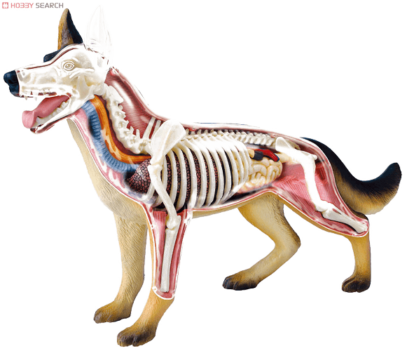
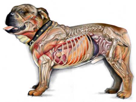
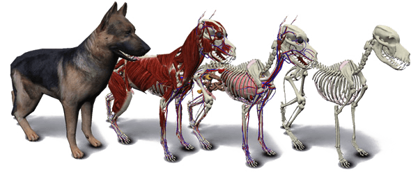
Dog Anatomy 3D Dog Skeleton 3D Dog Anatomy Free 3D Dog Anatomy Atlas Printable Dog Anatomy Charts Dog Bones Dog Eye Anatomy Dog Ear Anatomy Dog Anatomy Guide Dog Anatomy Tutorial Download Dog Anatomy Software Dog Head Anatomy Dog Paw Anatomy Dog Teeth Anatomy Dog Tail Anatomy What is the most sensitive part of a dog's body? What organ is on the left side of a dog? How is dog anatomy different from human? How many muscles are in a dog? Dog Anatomy Female Dog Anatomy Male Dog Anatomy Drawing Dog Anatomy Tendons Dog Tongue Anatomy Dog Skeleton Photos Dog Anatomy Information Dog Muscular System Dog Digestive System Dog Thorax Dog Abdomen Dog Organs Dog Nerves Anatomy Dog Anatomy Infographics Dog vs Human Anatomy Dog vs Horse Anatomy Dog Nasal Anatomy Dog vs Cat Anatomy Dog Muscles Dog Scull
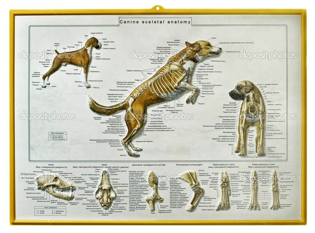
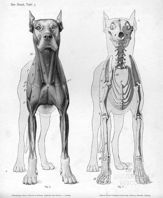
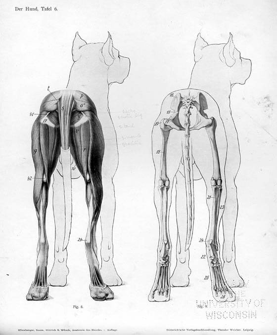
Dog has 320 bones in their body (depending on the length of their tail) and around 700 muscles.
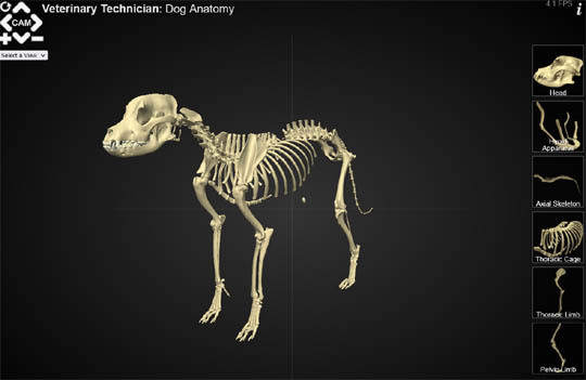
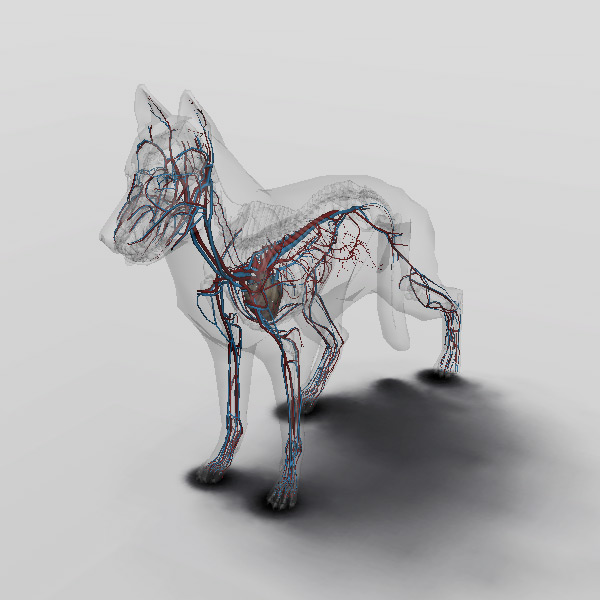
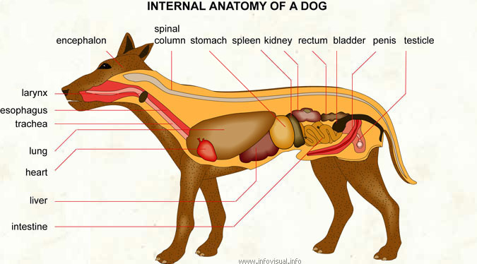
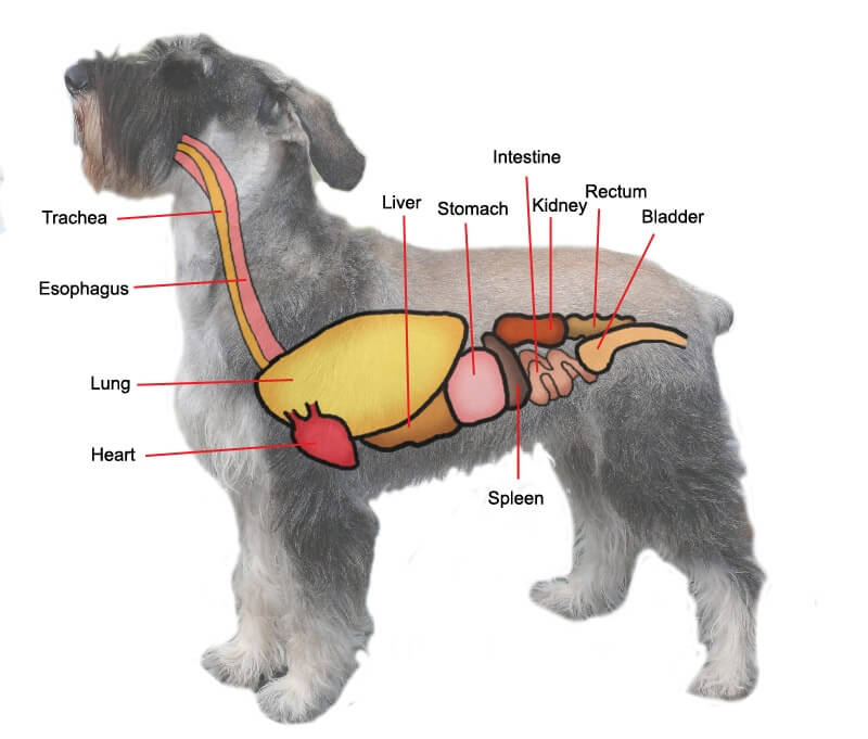
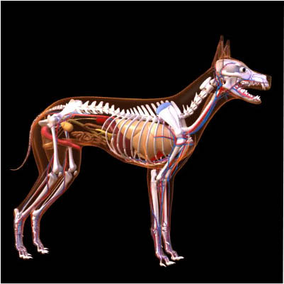
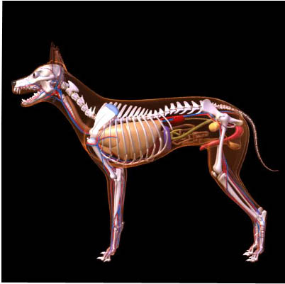
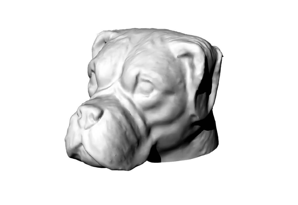
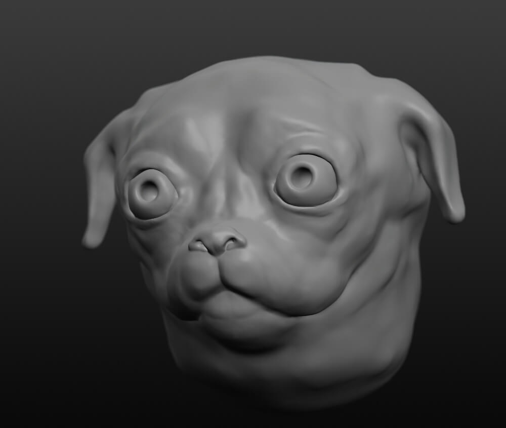
INTERACTIVE 3D DOG ANATOMY ONLINE This material proudly presented by WWW.DOGICA.COM
Delve into the dog body systems, peek under the skin and see what they are made of!
An immersive self-discovery experience into dog's body. You can manipulate bones, muscles, vessels, organs and other anatomical structures. Examine them closely from all angles, read their anatomical terminology.
This is also favored by the reflective membrane of the eye, the tapetum lucidum, which maximizes light when there's minimal light intensity. Moreover, the richness of rods in their eyes explain how dogs see in the dark, been able to walk through the house without tripping.
Another component of a retina is called a rod. Dogs have more rod cells than we do. Rod cells help see in dim light and also to distinguish the color gray.
Garcinia Cambogia Cute Little Fruit - It is believed that dogs can not only comprehend grey well, but that they can see many shades of grey that we cannot. Dogs, like cats, also see much better than we do in dim light situations.
Dogs are nearsighted creatures and do not see clearly at distances of more than 20 feet. They are able to detect movement at great distances, but cannot see the detail that would allow them to distinguish you from a small tree.
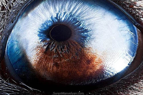
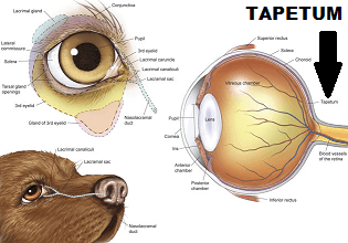
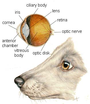
Dogs can see movement and light much better than people. In the retina of the eye, dogs have more of a specific type of cell called a rod, which is good at collecting dim light, so they have better night vision. A reflective layer in the dog's eye, called the tapetum lucidum, magnifies incoming light. This reflective layer lends a characteristic blue or greenish glint to dogs' eyes when light, for example, headlights of passing cars, shines into them at night. However, dogs do not have as much visual acuity as people, meaning that they cannot distinguish fine details as well. They also cannot differentiate colors as well because they have fewer of the cells in the retina called cones, which are responsible for color vision. Contrary to popular belief, however, dogs are not completely colorblind.
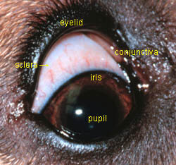
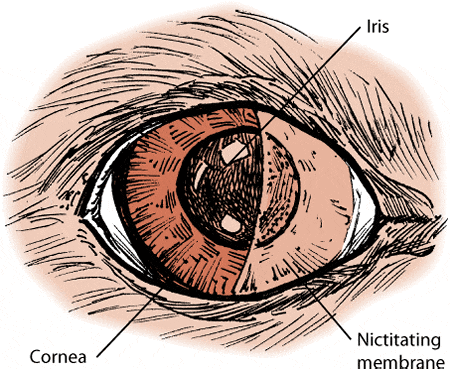
A unique feature of the dog eye is the nictitating membrane, which is also called the third eyelid. This additional eyelid is a whitish pink color, and it is found under the other eyelids in the inside corner (near the nose) of the eye. The third eyelid extends up when needed to protect the eyeball from scratches - for example, while traveling through brush or in response to inflammation.
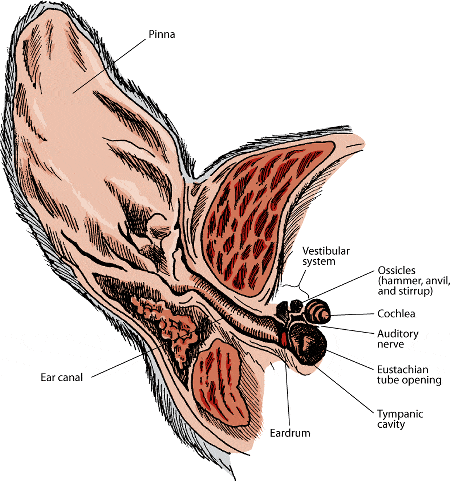
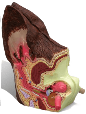
DOG EAR ANATOMY This article is proudly presented by
and
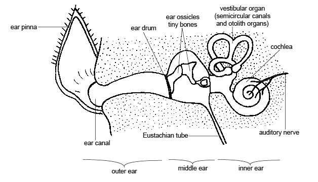
The ears are the paired receptor organs designed for the special senses of hearing and maintaining balance.
In many ways it could be said that a dog "leads with its ears." A dog's ears are right up front, one of the most noticeable parts of his anatomy, and they are a conspicuous visual reminder that demonstrates and carries much of his character and personality.
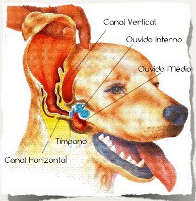
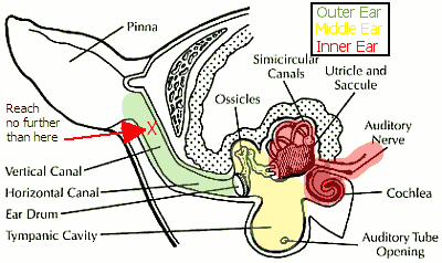
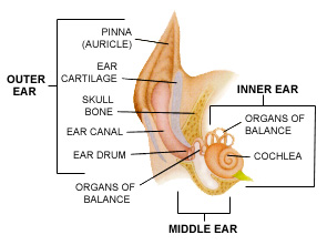
The ear is divided into three parts:
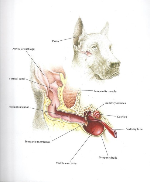
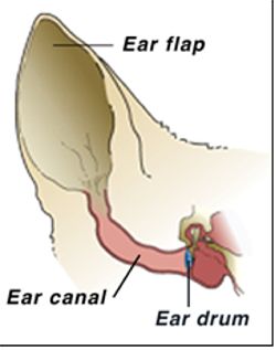
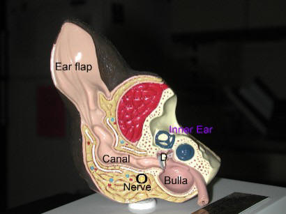
External (outer) Ear The external ear consists of the prominent earflap or pinna (also called the auricle) and the external ear canal (also called the auditory canal or meatus). The pinna is a funnel-shaped structure that collects sound and directs it into the external ear canal. The pinna is covered by skin, and the outer or posterior aspect is covered by fur. Numerous muscles are attached to the curved cartilage located between the inner and outer layers of skin around the ear, and these muscles allow the pinna to move and twitch. The external ear canal extends from the base of the pinna downward and inward towards the eardrum (also called the tympanic membrane). The external ear canal is L-shaped, with the L lying on its side. The canal forms an almost 90-degree angle between its two sections: the short, vertical outer section and the longer, horizontal inner section.
Middle Ear The middle ear includes the eardrum and the bony tympanic cavity (osseous bulla), which lies just past the ear drum. Within this tympanic cavity are found the auditory ossicles: three tiny bones that vibrate when stimulated by sound waves. These ossicles are named the malleus, stapes and incus, commonly known as the hammer, the stirrup and the anvil because of their resemblance to these objects. These three bones form a chain across the middle ear from the tympanum to the oval window of the inner ear. The middle ear is connected to the back of the throat (pharynx) by the auditory or eustachian tube. This tube allows air from the pharynx to pass in and out of the middle ear, which helps keep middle ear pressure normal. The middle ear is connected to the inner ear through the oval window, which lies against the stapes bone.
Internal (inner) Ear The inner ear is located within the petrous temporal bone of the skull and consists of two parts. The osseous or bony labyrinth houses a series of thin, fluid filled membranes called the labyrinth. The inner ear contains three distinct structures: the cochlea - spiral tube, vestibule, and three semicircular canals. The cochlea contains the nerves that transmit the electrical impulses and is directly responsible for hearing. The vestibule and semicircular canals are responsible for maintaining balance or equilibrium. These tissues are supplied by the two branches of the 8th cranial nerve - the vestiblocochlear nerve, which transmits electrical impulses related to sound and balance back to the brain.More than a dozen separate muscles control the movement of the ear, and the entire area is richly supplied with blood vessels and nerves.
The ear has two functions: Hearing and Balance - and either function can be disturbed by disease, old age, or nerve disruption from a number of causes.
Hearing. Hearing is one of the keenest senses in dogs, together with smell. Sound first enters the external ear canal as sound waves. As these waves strike the eardrum, it begins to vibrate. These vibrations are then transmitted to the three small bones of the middle ear - the malleus, incus and stapes, which amplify the sound vibration. The end of the stapes is connected to the oval window of the inner ear. As the stapes vibrates, it transmits the sound vibrations to the cochlea, the snail shaped portion of the inner ear, which transforms the vibrations into nerve signals that are transmitted to the brain where they are interpreted as sound.
What dog hears? Hearing can be visualized as waves of energy traveling along molecules in the air, transformed into mechanical energy at the ear drum, then amplified by small bones and finally transformed into the electrical impulses in the auditory nerve, resulting in what the brain registers as hearing.
Dogs have a much different range of hearing than ours, extending into a considerably higher frequency than we can hear. Sound frequency, the number of sound wave cycles every second, is measured in Hertz (Hz). The higher the frequency, the more sound waves per second, the higher-pitched the sound. Humans hear best at around 2,000 Hz; dogs hear best at 8,000 Hz – perhaps the reason they respond better to high pitched cues.
Balance. The other function of the ear is to help maintain balance. The three semicircular canals of the inner ear are oriented at right angles to each other. When the head turns, the resulting movement of fluid in these canals allows the brain to detect which way and how much the head is turning. Another part of the inner ear responds to gravity and sends information to the brain when the head is held still in a stationary position.
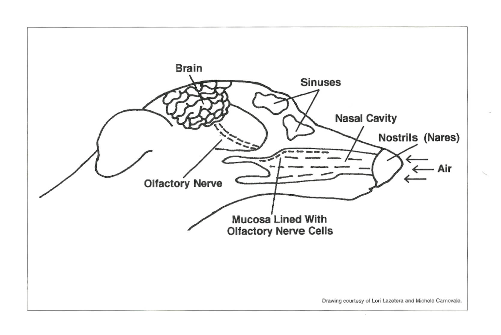
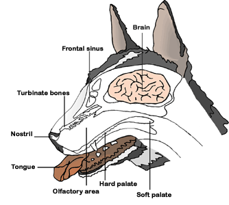
DOG NASAL ANATOMY This article proudly presented by WWW.NATURAL DOGCOMPANY.COM and WWW.PREZI.COM and WWW.DOGICA.COM
In case you have not noticed: your dog's world revolves around his nose.
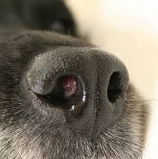
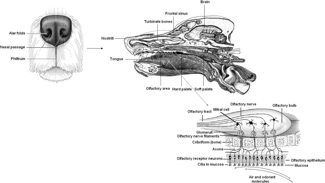
Dog's noses are not like our noses. They are so much more! In fact, a dog uses its nose much like humans use their hands. Dog's noses are highly sensitive and when those tissues are dry and chapped, it can be very painful to your pup.
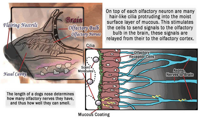
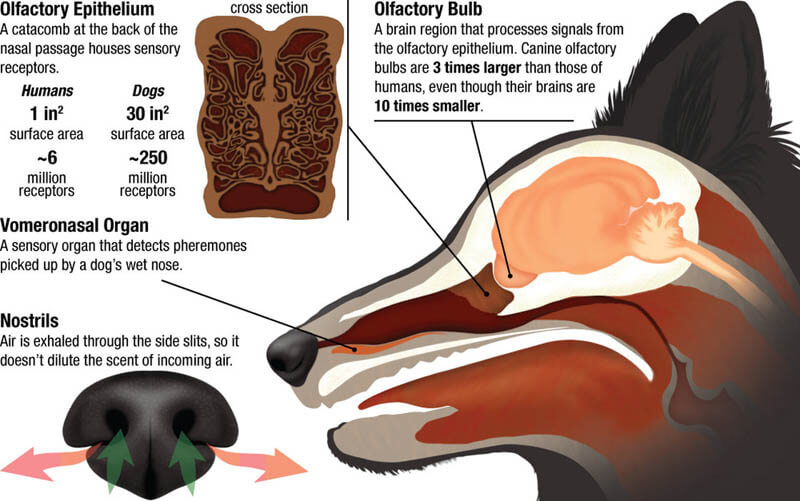
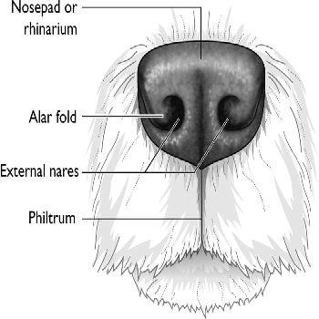
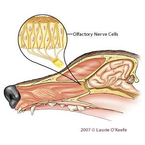
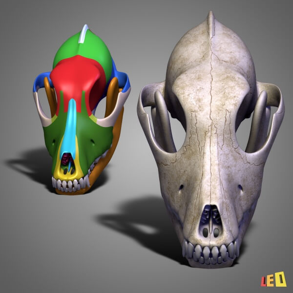
A dog's nose consists of a nasal cavity and a pair of nostrils (nares) for inhaling air and odors. The olfactory receptor cells in a dog's nose extend throughout the entire layer of specialized olfactory epithelium found on the ethmo-turbinate bones of the nasal cavity. The olfactory portion of the nasal mucous membrane contains a rich supply of olfactory nerves that ultimately connect with the highly developed olfactory lobe in the dog's brain.
Dogs possess an additional olfactory chamber called the vomeronasal organ that also contains olfactory epithelium. The vomeronasal organ, known as Jacobson's organ, consists of a pair of elongated, fluid-filled sacs that open into either the mouth or the nose. It is located above the roof of the mouth and behind the upper incisors. Interestingly, the olfactory receptors in the nasal cavity are anatomically distinct from those in the vomeronasal organ.
Each receptor neuron (nerve cell) in the olfactory epithelium of the nasal cavity has a dendrite that ends in a knob with several thin cilia covered by mucus. Receptor neurons in the vomeronasal organ typically lack cilia but have microvilli on the cell surface.
A dog's nose should be cool and moist. A dry and or warm nose indicates an unhealthy and uncomfortable nose. The moisture secreted by mucous glands in the nasal cavity captures and dissolves molecules in the air and brings them into contact with the specialized olfactory epithelium inside the nose.
Dogs use sniffing to maximize detection of odors. The sniff is actually a disruption of the normal breathing pattern. Sniffing is accomplished through a series of rapid, short inhalations and exhalations. A bony subethmoidal shelf, which is found below the ethmo-turbinate bones of the nasal cavity, forces inhaled air into the olfactory epithelium. Washing out of the region upon exhalation does not occur due to the nasal pocket created by the bony subethmoidal shelf.
The nasal pocket permits the odor molecules that are unrecognizable in a single sniff to accumulate and interact with olfactory receptors. Odor molecules in the olfactory epithelium of the nasal cavity are absorbed into the mucous layer and diffuse to the cilia of receptor neurons. This interaction generates nerve impulses that are transmitted by the olfactory nerves to the dog's brain, which has a well-developed olfactory lobe. This allows the dog to recognize a scent and follow a trail.
Olfactory receptor cells in the vomeronasal organ also send impulses to the region of the hypothalamus associated with sexual and social behaviors. This organ is believed to be important in the detection of pheromones (body scents). This theory could account for the dog's ability to identify and recognize other animals and people.
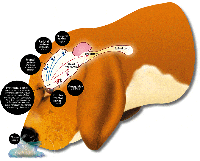
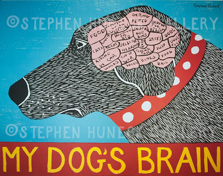
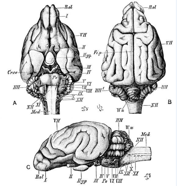
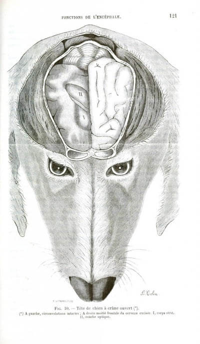
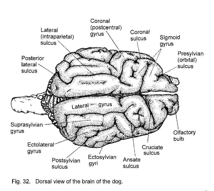
DOG BRAIN ANATOMY This material proudly presented by WWW.EHOW.CO.UK
3 MAIN AREAS OF CANINE BRAIN
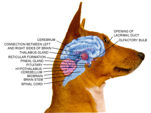
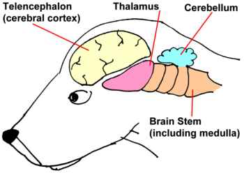
Telencephalon The front part of the brain is called the telencephalon. Information from the five senses is interpreted there, and it is also where thought occurs. Dogs have large telencephalons which makes their ears, nose and eyes exceptionally sensitive. It also is responsible for dogs' undeniable personalities, and their advanced social behaviours.
Diencephalon Behind the telencephalon lies the diencephalon. Most basic functions are controlled in this portion of the brain. Chewing, breathing, equilibrium and the collection of information from the senses all occur here. This part of the brain is highly advanced in dogs, contributing to their fast reflexes, agility and the acuteness of their hearing.
Metencephalon This part of the brain is behind the diencephalon. It is responsible for finer muscle skills and the regulation of blood flow and pulse rate, and is also the brain's reward centre. For dogs, this part of the brain contributes to their remarkable endurance and stamina and is the part of the brain responsible for their love of playing fetch and other games.
Medulla Oblongata At the base of a dog's brain, where it connects to the spinal cord, is a structure known as the medulla oblongata. Here the basic functions that occur without thinking are regulated. Digestion, heart beat, respiration, swallowing and sneezing are all controlled in this area of the brain. The medulla oblongata is the first part of the brain that develops in puppies before they are born.
Corpus Callosum In the middle of a dog's brain is the corpus callosum. This is a wall of nerve cells which facilitates communication between the left and right side of the telencephalon and diencephalon. Depending on the breed of dog, the corpus callosum's size and the speed at which it allows the halves of the brain to interact can vary significantly.
As mammals the dog's brain has a fairly similar structure to the human brain: The cerebral cortex is the part of the brain responsible for the way in which we use our senses, such as sight, touch, vision and taste. The cerebral cortex of the dog has pronounced ridges on the surface, and this implies how advanced their brain is at distinguishing between these sensory stimuli. Dogs need this finely tuned hearing and sight because they have to hunt in order to survive, as well as protecting themselves and their pack.
Dogs are famous for their impressive sense of smell. The olfactory bulb is responsible for this and in dogs this area is very large, 150cm squared compared to 5cm squared in humans. Sense of smell is vital. They use it to interpret the world around them and everything within it. Just think how much we use our vision to find our way around the world - to a dog, his sense of smell is just as important. We've been able to utilise this in order for the dog to help us: search and rescue and sniffer dogs are now vital and enable us to live in a much safer society than we would be able to otherwise thanks to a nose 1,000 times more powerful than our own.
The temporal lobes on the canine brain are also very pronounced. This is the section where memories are stored. This will not come as a surprise to many dog owners - when we take our dogs to their favourite field where they seem to remember chasing a rabbit many months ago, even if they haven't been there for a long time. This supports the idea that dogs can have an impressive memory; vital for canines and wolves living in the wild. Perhaps this part of the brain has evolved in this way because a good memory is vital to their long term survival.
The frontal lobes are situated in the cerebral cortex and are famous for being responsible for our human intelligence. We are the most intelligent species on earth - we invent new medicines and discover the complexities of space. Without this vital section of the brain none of this would be possible. All species have a frontal lobe they just are not as developed as our own. The frontal lobes of the canine brain are fairly long compared to many smaller mammals. This part of the brain is responsible for the dog's intelligence and its nature. Many breeds are bred specifically for their intelligence and have been for generations; Border Collies for example are widely considered to be the most intelligent of all the breeds and therefore need a huge amount of mental and physical stimulation.
The complexity of the appearance of the canine brain, hints at the intelligence of the dog, and the way in which it has evolved over many centuries. It will be interesting to see what the brains of these animals look like in another 500 years and how they will further evolve to adapt to the world in which they live.
To be more knowledgeable in the area of canine brain read the following articles:
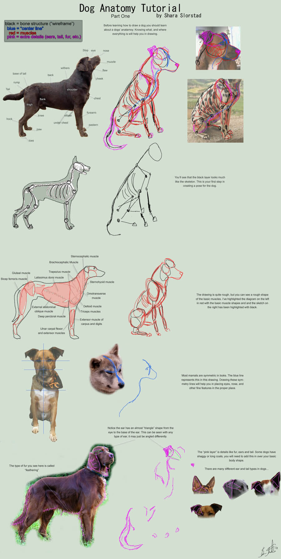
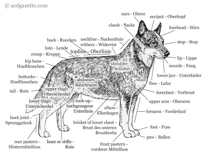
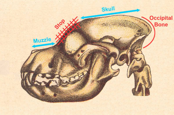
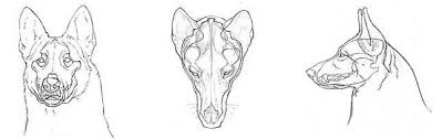
Dogs' heads have 5 different components each of which can vary considerably. Some variations of the components of dogs' heads are distinctive whilst others are quite subtle. So it is imperative to understand the different components that make up the head before we can discuss these variations. After all, the dog's head sets the breed type of each particular pure breed of dog.
Starting from the head, a dog is made up of the
Nose: Dog noses are often cold and wet, and of course, they usually get stuck where they're not wanted.
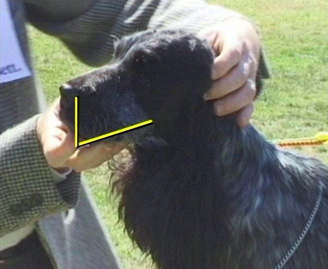
The muzzle (foreface) comprised of the upper and lower jaws. The foreface or muzzle is the whole of the upper area from the eyes to the nose including the lips. It is sometimes also referred to as the face.
The boney part on the top of the muzzle is often called the nazal bridge whereas the sides of the muzzle are often referred to as flews in Breed Standards and general dog jargon. Some Breed Standards refer to the flews as muzzle. So, confusion could arise as to what are flews and what exactly is the muzzle. For example, the Breed Standard of the Cocker Spaniel shown here says it should have a square muzzle, whereby it is obvious that it is the flews that are square shaped.
The boney part on the top of the muzzle is often called the nazal bridge whereas the sides of the muzzle are often referred to as flews in Breed Standards and general dog jargon. Some Breed Standards refer to the flews as muzzle. So, confusion could arise as to what are flews and what exactly is the muzzle.
For example, the Breed Standard of the Cocker Spaniel shown here says it should have a square muzzle, whereby it is obvious that it is the flews that are square shaped. When the muzzle or flews look thick or padded, this is called cushioning. For example the Tibetan Spaniel shown here. This cushioning gives the dog a soft expression. Technically the lower lips are that potion of the skin closest to the mouth cavilty that is devoid of hair. The lower lips blend into the skin of the chin while upper lips blend into the skin that covers the top teeth.
The stop is an indentation (sometimes nonexistent) between the muzzle and the braincase or forehead.
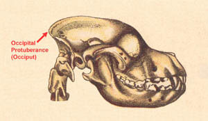
The Occiput is the highest point of the skull at the back of the head and a prominent feature on some dogs.
The occipital bone has a crest or protuberance which is commonly referred to as the occiput. It is is clearly seen here in the English Setter and Bloodhound. However, in some other breeds it is barely perceptible. Myths in dog folklore believed that size of the occiputal protuberance was a measure of the dog's sense of smell. So to this day it is prominent in most Scent Hounds.
But the occipital bone itself actually extends right down the back of the head to where it articulates with the neck. So when breed standards refer to the length of a dog's skull, this measurement does not include the occiput as this is part of the occipital bone.
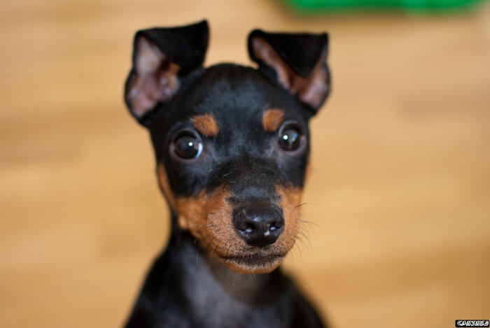
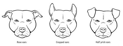
It's well known what ears are, but different dogs have different types of ears, including:
Pricked: Pricked ears are upright.
Dropped: Dropped ears hang down.
Button: Button ears have a fold in them.
Cropped: Cropped ears are surgically altered.
Eyes are pretty obvious, and most often obviously brown.
Like humans, dogs have eyebrows, or simply brows.
Whiskers provide some sensory feeling.
Flews is just a fancy word for a dog's lips.
A dog's cheek is the skin along the sides of the muzzle, about where your cheeks are if you had a muzzle.
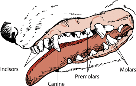
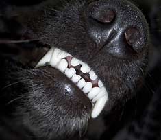
Dogs have 42 teeth. Six pairs of sharp incisor teeth are in front of the mouth, flanked by two pairs of large canine ("dog") teeth. The other teeth are premolars and molars. The incisors and the canines are very important because the dog bites and tears at its food with these teeth.
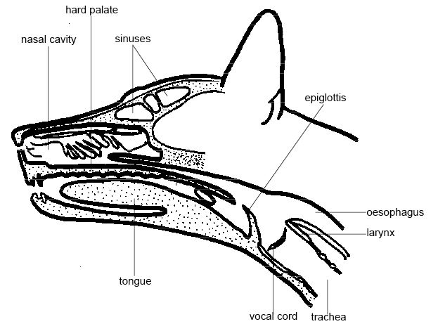
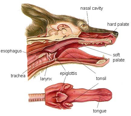
Air breathed in through the dog's nose passes on its way to the lungs through the two nasal cavities behind the nose. These cavities are lined by a mucous membrane containing many nerve endings stimulated by odors. Smell is the dog's most acute sense.
A dog continually sniffs the air, the ground, and nearby objects to learn what is happening around it. The indentation in the dog's forehead just above eye level is called the stop. The stop in some dogs is deeper than that in others.
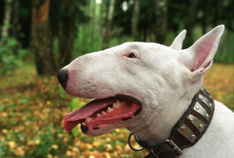
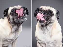
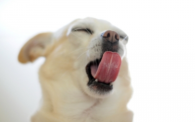
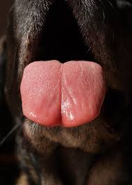
The fairly thin tongue of the dog is used mainly for guiding food to the throat, for licking the coat clean, and for perspiration. When a dog is overheated, it cools off by hanging its tongue out and panting. As it pants, the evaporation of perspiration from its tongue cools the animal. The dog also sweats through the pads on its paws and slightly, through its skin.
A dog's ears either stick up or hang down. The earliest dogs probably had erect ears, but the ears began to droop in smaller, later breeds because of excessive ear skin. Dogs have a fine sense of hearing. They can hear sounds at frequencies too high for people to hear. This is why dogs can respond to "silent" whistles.
Each eye of a dog has three eyelids, the main upper and lower lids and a third lid hidden between them in the inner corner of the eye. The third eyelid can sweep across the transparent cornea of the eye and clean it like a windshield wiper.
The head and body of a dog are connected by its neck. The neck may be long or short, depending on the size of the seven bones that support it. The length of the vocal cords in the neck is a factor influencing the pitch and loudness of a dog's voice its barks, grunts, and howls.
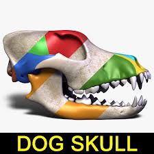
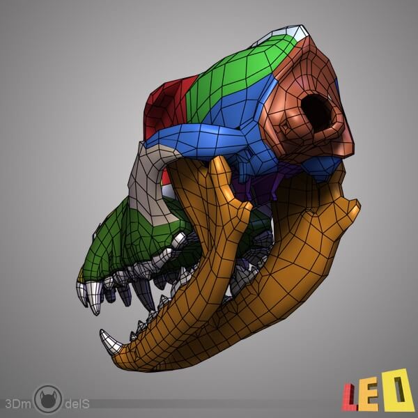
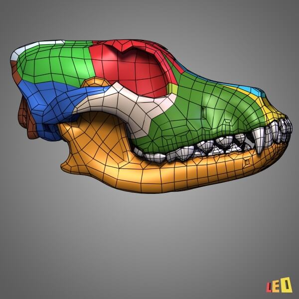
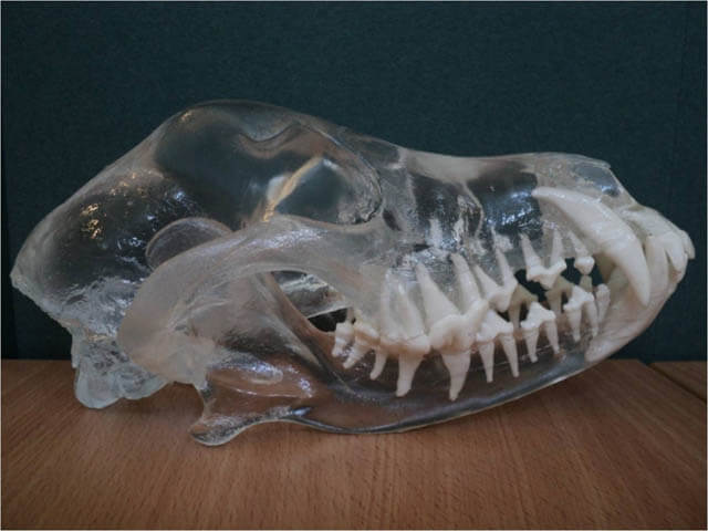
DOG HEAD ANATOMY (Skull) This article proudly presented by WWW.REAL3D ANATOMY.COM and WWW.3DMUSEUM.ORG
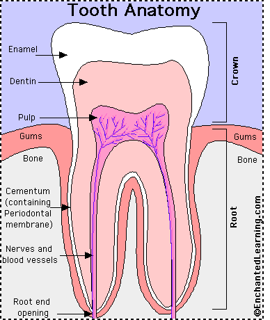
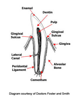
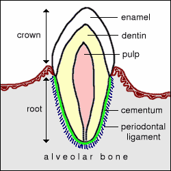
Teeth are a set of highly mineralized living tissues used by mammals to hold, tear, and chew. They are significant not only for eating, but also for protection. The teeth even play an important role in keeping the tongue safely moist inside the mouth.
Each tooth has a crown (located above the gums) and a root (located below the gums). Some teeth, such as incisors, have one root, while others, such as the largest cutting premolar, called the "carnassial tooth," has as many as three roots.
A dog tooth is composed of the following structures:
Pulp: The pulp is at the center, or core of the tooth, and consists of connective tissue, nerves, and blood vessels that nourish the tooth. Most of the nerves and blood vessels to the tooth enter through the apex (bottom) of the root. Special cells in the pulp, called "odontoblasts" form dentin.
Dentin: The majority of the tooth is made up of dentin, which surrounds the pulp. Dentin is as hard as bone but softer than enamel. Dentin is a tissue that can detect touch, heat, and cold. Primary dentin is dentin that is formed before tooth eruption, secondary dentin is dentin that is continually formed throughout the life of the tooth. As the secondary dentin forms, the pulp chamber reduces in size. The dentin of the crown is encased in enamel and the dentin of the root is covered by cementum.
Enamel: Enamel is the hardest tissue in the mammalian body and is formed before tooth eruption. Just before the tooth erupts through the gums, the formation of enamel stops and is lost gradually over the life of the tooth. Although enamel is very hard, it is brittle, too, often subject to chipping. The tissues that surround the teeth are called the "peridontium" and consist of the alveolar bone, periodontal ligaments, cementum, and gingiva.
Alveolar Bone:The alveolar bone forms the jaw and the sockets into which the roots of the teeth extend.
Periodontal ligaments: This tough tissue helps to hold the tooth in the socket. It attaches to the cementum of the tooth and the alveolar bone.
Cementum: Cementum is hard, calcified tissue that covers the dentin of the root and is slowly formed throughout the life of the tooth. It assists in supporting the tooth in the jaw and in root repair.
Gingiva: The gingiva, also called the "gums," is the soft tissue that covers the rest of the peridontium.
Lateral canal: The lateral canal is a very small channel that connects the root pulp to the periodontal tissue through which small blood vessels run.
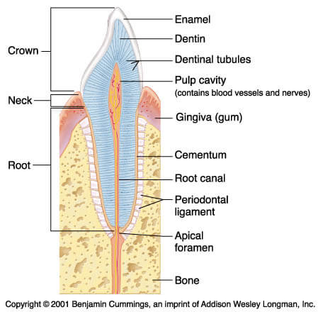
Although teeth have different shapes and functions, each still shares the same structural anatomy. The innermost portion is the pulp/root canal, in which the blood vessels and nerves lie. Surrounding this canal, the dentin provides the structure for most of the tooth. The shiny, protective enamel covers the outer part of the crown, which is the visible portion of the tooth. The roots of the tooth are firmly held into the jawbone with the periodontal ligament. This strong sheath of tissue "glues" the tooth root to the surrounding bone. The gum tissue overlying the base of teeth is called the gingiva.
Teeth are very important to an animal as they are used for eating, grooming and defense. Consequently, dental problems, if not treated, often lead to more generalized illness. Mammals have teeth of different sizes and shapes, a condition known as heterodonty, allowing different teeth to be specialized for different tasks.
These specialized teeth include: Incisors, Canine teeth, Premolars, Molars. Mammals also have two sets of teeth: a deciduous set (milk teeth, baby teeth) and a permanent set.
In mammals, there are two distictive types of teeth that differ in pattern of growth and morphology:
Brachydont or low-crowned teeth are what is seen in man, carnivores such as dogs and cats, and pigs. This type of tooth consists of a crown above the gingiva, a constricted neck at the gum line, and a root embedded in the jawbone. The crown is encased in enamel and the root in cementum. Enamel is the hardest substance in the body being densely packed with hydroxyapatite mineral crystal and heavily mineralized with calcium salts. Cementum is calcified connective tissue. Dentin, a bonelike material, is under the enamel and makes up most of the tooth. The pulp cavity includes blood vessels, lymphatics and nerves.
Hypsodont or high-crowned teeth are continue to erupt throughout life. Examples of this type of teeth include all of the permanent teeth of horses and cheek teeth of ruminants. Hypsodont teeth are usually described as having a body, much of which is below the gum line, and root, which is embedded in the alveolus of the jaw bone. Enamel covers the entire body of the tooth, but not the root.
In both high-crowned and low-crowned teeth, the tooth is attached to a "socket" in the jaw bone called an alveolus. The attachment is through a fibrous capsule called the gomphosis. Remembering the term gomphosis is required only of dental students. An additional type of nomenclature is used to describe the different surfaces of teeth, as depicted in the image below. The occlusal surface is the chewing surface.
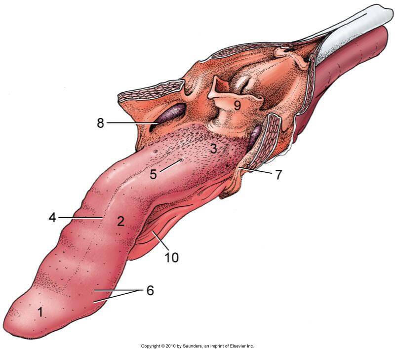
Dog's tongue fed by five separate sets of nerves that come directly from the brain through small openings in the skull. These are cranial nerves, and they originate from the base of the brain rather than from the spinal cord.
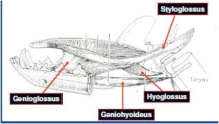
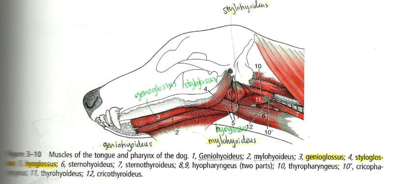
The tongue is made up of 6 major muscles, the Frenulum, which attaches the tongue to the floor of the mouth, the Genioglossus, which pulls the tip of the tongue back, the Hyoglossus, which draws the tongue back into the mouth, the Styoglossus, which pulls the tongue back and upwards, and the Mylohyoideus, which supports the extrinsic muscles of the tongue.
Dogs depend on their sense of taste to find resources like food and water.
The dog's taste buds are as follows:
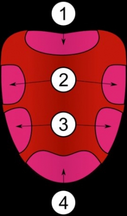
1. Salty 2 & 3.Sweet 4. Water - yes water!
Dogs have a finely tuned ability to taste water, which comes in handy when looking for something to drink.
Dogs taste sour over much of the top of the tongue towards the back with sweet on the sides and the front. If you use taste deterrents such as Bitter Apple to prevent your dog from chewing inappropriate items like your shoes, I find it helpful to spray it in the dogs mouth first so they associate the product in the bottle with the bad taste, then let them see you spraying it on the forbidden item. Dogs make negative taste associations quickly, so this usually does the trick and they will know the item tastes like whatever is in that yucky bottle. The reason why it is not helpful to spray the item before the dog has tasted it is because when a dog chews, it is usually the sides and front of their tongue that come in contact with the object, not their bitter taste buds, so they do not taste the taste deterrent (I know, weird right?).
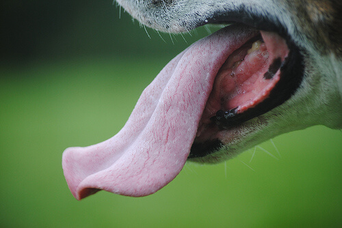
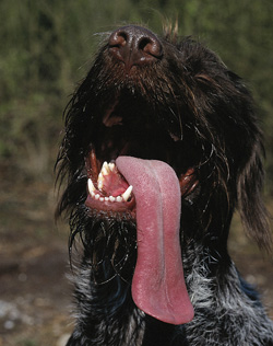
Because dogs cannot sweat they depend on their tongue as a major source of heat loss. The tongue is rich with capillaries so when a dog pants, the tongue swells and the rapid movement of cool air from the environment moving over their moist tongue whisks away heat, helping them regulate their body temperature.
One of a dog owner's most enjoyable moments is watching their dog running and playing with their tongue flapping in the breeze. While tongue injuries are rare, they can happen, especially if they are running in tall grass where foxtails can get caught in their mouth. These need to be removed immediately to prevent them from becoming embedded and infected. Cuts on the tongue can also happen, and because the tongue has such a large amount of capillaries, small lacerations can bleed quite profusely. In this case, it looks much worse than it actually is, so if the dog is panting and the tongue is swelled, cooling the dog down will reduce the swelling and allow the tongue to clot quickly and the bleeding will stop.
The tongue is a remarkable organ, but we as dog owners tend to love it more for the kisses it gives us! We tend to look at big wet sloppy puppy kisses as a sign of affection from our dogs, but is it? As puppies, a mother dog uses licking as a way to keep her pups clean, to stimulate them to urinate or defecate, and to encourage bonding between her and her pups. Licking also helps stimulate their mental development. As the puppies grow, usually after 6 weeks of age, puppies begin to return the favor and will lick their mother's lips when they want her to regurgitate food for them to eat. Licking is also a sign of submission that is used in dog-dog interactions, as well as a part of grooming. Dogs will also lick when they are nervous or as a gesture of appeasement or goodwill.
In an article by Dr. Nicholas Dodman, dogs "may lick their own lips or may lick a person to whom they wish to signal deference. If the recipient of the licking interprets this behavior as "make-up kisses," that's just fine. Perhaps the behavior is analogous to some forms of human kissing and thus their interpretation may be close to the truth." But what about that excited dog that jumps all over us when we get home and licks our faces with reckless abandon? Dodman explains, "For some dogs, it seems that they engage in face licking because they can get away with it and because it gets a rise out of the person." This might be a case of positive reinforcement where 1. dog licks person, 2. person gets excited and rewards the dog with petting, praise and affection.
Lesson learned - giving kisses is good!
Whether you are a fan of "getting kisses" from your dog or not, or whether you think your dog is simply begging to share what you had for lunch or is giving you genuine affection, we can all agree that the tongue is of vital importance to our dogs. The tongue is a sustainer of life, an air conditioner, a bath, a former of bonds and a great communicator. The tongue is a muscle that really pulls its weight!
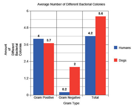
Dogs secrete saliva which contains an enzyme that kills certain bacteria known as lysozyme therefore it is regarded as having antiseptic tongue. However, the cleanliness of its mouth depends on the specific dog and its activities.
It's a radiator, a water lapper, a healer of wounds, a food conveyor, a register of tastes, a texture sensor, and a wet equivalent of a dog's handshake. A dog's tongue has more responsibilities than any other part of the dog anatomy excluding the brain. And oddly enough, for all its duties and actions, it is one of the most maintenance free structures of all the dog's body parts!
Let's take a look at unique structure & see what we can discover: On a recent photo shoot with one of my dog trainer/hunter friends, I exposed four rolls of film while he put this three black labs through some off-season training. When I placed the slides on the viewer I was curiously struck by how many action shots captured the charging subjects with their long, flexible tongues literally flopping out there in the breeze. (I'm talking about the dogs here, not the trainer!)
Almost every photo displayed the dog's tongue completely extended with mouth open wide, fully exposing the airway to the onrushing breeze. After seeing these photos, I was amazed that in my busy small animal practice I wasn't seeing more than just occasional tongue injuries. With that fleshy, vascular flag waving around, frequent injuries should be expected, but in 25 years of practice in an area pleasantly infested with hunting dogs, tongue problems are just not very common.
Nevertheless, it has happened more than a few times that I would get a frantic call at home from a hunter wanting to rush his gun dog in because "she's bleeding from the mouth like a stuck pig!" So I'd rush in to the animal hospital expecting to perform some heroic surgery only to find the bleeding had stopped and the owner apologetic about all the fuss. Upon examining the mouth, I'd find one or more lacerations: sometimes not very substantial at all - that had clotted and nicely sealed.
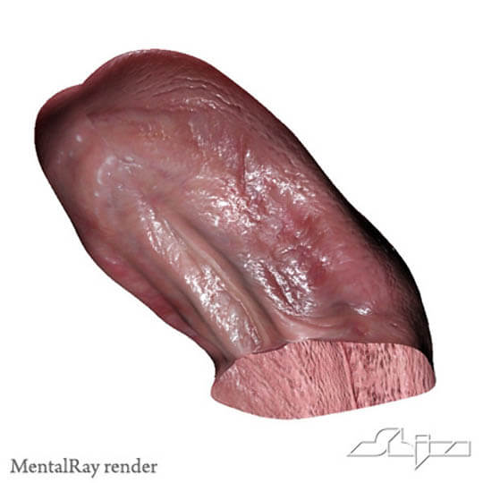
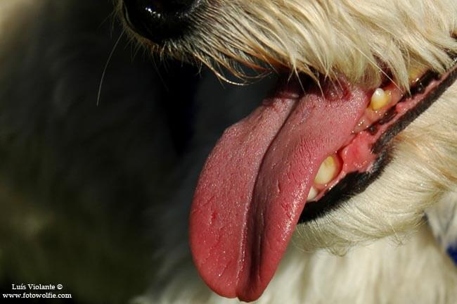
Examining the Tongue's Anatomy Essentially the tongue is an elongated muscular organ with the top surface covered with specialized epithelium. Its responsibilities include responding to taste, touch, pain, and aiding in heat dissipation.
When I began researching this article, I quizzed myself and was able to recall only three muscle groups interacting with the tongue. Well, the faithful Miller's Anatomy of the Dog describes no less than eight pairs of muscles whose job it is to control the tongue's activities. They have intimidating Latin names such as genioglossus vertical and oblique, hyoepiglottis, and sternohyoideus.
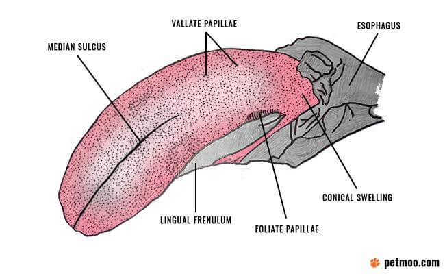
That band of tissue directly under the tongue holding it down, that's called the frenulum, you've got a frenulum too, only not quite so well developed.
And something you don't have that the dog does: feel just under the tip of the dog's tongue running from front to back along the midline, you'll find a firm cartilaginous, almost bony structure. That's called a lyssa. This little device was considered in ancient times to be a cure for various ailments including rabies!
TASTE: In addition to directing the dog to eat rotten garbage and to be repulsed at the taste of woodcock, the canine tongue is capable of discerning sensations of salt, sweet and sour. The sensation of sour is dispersed somewhat evenly over the top of the tongue, salt along the lateral edges and rear of the tongue and sweet along the edges and front of the tongue. Dogs have a finely tuned ability to taste water, and that trick is performed only by the tip of the tongue.
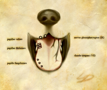
PAPILLAE: These odd projections from the surface of the tongue are of five different types. The slightly shredded look to the front and side of the dog's tongue (especially noticeable in newborn pups) are called marginal papillae and those funny bumpy things on the back of the tongue are vallate. Now the next time you see your buddy curiously peering into his dog's mouth and he suddenly exclaims, "Hey, what the heck are these weird doofangles on Cinder's tongue?", you can tell him they're called papillae and there are five kinds of them and casually walk away.
DOG TONGUE INFECTIONS: Because it is so richly supplied by nourishing blood vessels, infections of the tongue are not common. Generally, when they do occur, a foreign body such as a fox tail awn, porky quill, thorn or wood splinter is the culprit and can be removed under anesthesia. (Anyone who lets their dog chew on lumber, please stand up, uh huh. Okay, everybody can sit down now.) Split firewood and 2x4's sure can make a dog proud and happy, but those woody splinters can wreak havoc in the dog's mouth and gastrointestinal tract. Wood is indigestible, you know. Throw them a tennis ball and forget the timber!
It's a good idea to really examine your dog's mouth routinely say every Saturday morning just before you start on those chores you've been putting off. Maybe if you're lucky, you'll find something suspicious requiring an immediate trip to the animal hospital and thereby a legitimate postponement of the chores until the following Saturday!
WIRING: The canine tongue is uniquely constructed to do so many things. And to perform all these diverse and intricate functions the tongue requires five separate pairs of nerves coming directly from the brain through tiny openings in the dog's skull. These are called Cranial Nerves since they do not arise from the spinal cord, but directly from the base of the brain itself. In many an idle moment I have pondered what effect on my shooting success there would be if I had a fancy cranial nerve connected to my right forefinger rather than an ordinary spinal nerve.
Remember, the tongue is king!
Everything else in the mouth is an assistant. Keep a close watch, though, for ulcers, bruises or bleeding from the tongue, gums or palate. Check for broken teeth that can irritate the tongue or bumps arising anywhere within the oral cavity. Work your finger under each side of the tongue and force it upward so you can inspect the underside of the tongue. I've found some pretty odd things wedged or otherwise hiding beneath the tongue. You really should reward that tongue once in while by allowing it a full, wet slap on your face just before its owner bounds off on a walk with you just for fun - no dummies, no whistles, no check cords or leashes. Odds are that the tongue will reward you at the end of your playful excursion.
ANATOMY OF DOG's TAIL This article proudly presented by WWW.DOGICA.COM
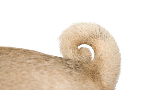
A dog's tail is the end of his spinal column. It is attached to the backbone, though attached isn't exactly the right word. In fact, a dog's tail is the hindmost end of his backbone. It consists of six to 23 vertebrae enclosed by muscles that are attached to the vertebrae by tendons. The highly mobile and flexible vertebrae and muscles give the dog an enormous amount of control over how his tail moves. He can lift it, move it from side to side or pull it down to cover his anus and tuck between his legs. In every one of these positions, the tail is capable of many different movements, making it nearly as expressive of emotions as a human's face.
A dog's tail is an extension of the spine. It helps them to keep balance when they run. Moreover, A dog's tail position and motion is incorporated as a component of a complex system of body language that domestic dogs use to show excitement or agitation.
The caudal muscles lie on the lumbar vertebrae, sacrum (in the lower back region) and tail vertebrae. The muscles insert on the tail/caudal vertebrae exclusively. The muscles are attached to the tail vertebrae by tendons. The most posterior tendons attach to the last tail vertebrae.
Part of the musculature is formed from muscles associated with the rectum, the anus and the pelvic diaphragm. Four to seven paired nerves serve the tail muscles. These muscles have many tendons that insert from the fifth or sixth caudal vertebra, then onto the next vertebra, and so on to the end of the tail.
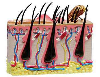
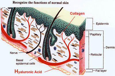
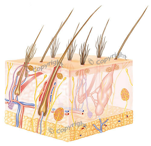
The skin is the largest organ of your dog's body. It provides a protective barrier against the environment, regulates temperature, and gives your dog its sense of touch. Depending on the species and age, the skin may be 12 to 24% of a dog's body weight. The skin has 3 major layers: the epidermis or outermost layer, the dermis or middle layer, and subcutis or innermost layer. Other important parts of the skin include skin appendages (such as hair and claws) and subcutaneous muscles and fat.
The anatomy of a dog's skin includes 3 major layers, as well as hair follicles and sebaceous glands.
Epidermis is the outer layer of skin. It provides protection from foreign substances. The epidermis is composed of multiple types of cells, including keratin-ocytes, melanocytes, Langerhans cells, and Merkel cells. Each of these cells has special functions.
Keratinocytes provide a protective layer that is constantly being renewed in a process called keratinization. In this process, new skin cells are created near the base of the epidermis and migrate upwards. This produces a compact layer of dead cells on the skin surface. This layer keeps in fluids, salts, and nutrients, while keeping out infectious or noxious agents. The top layer of dead skin cells are continuously shed and replaced by cells from lower layers. The rate of cell replacement is affected by nutrition, hormones, tissue factors, immune cells in the skin, and genetics. Disease and inflammation also alter normal cell growth and keratinization.
Melanocytes are located at the base of the epidermis, the outer root sheath of hairs, and the ducts of the sebaceous and sweat glands. The melanocytes produce the skin and hair coloring (pigment) called melanin. Production of melanin is controlled by both hormones and the genes received from parents. Melanin helps protect the cells from the damaging rays of the sun.
Langerhans cells are part of the immune system. These cells are damaged when exposed to excessive ultraviolet light and glucocorticoids (anti-inflammatory drugs). Langerhans cells play an important role in the skin's response to foreign substances and contribute to such things as the development of rashes when an animal is exposed to irritating materials.
Merkel cells are specialized cells associated with the sensory organs in the skin. In particular, Merkel cells help provide animals with sensory information from whiskers and the deep skin areas called tylotrich pads.
Basement Membrane Zone This layer of the skin is located beneath the epidermis and connects the epidermis to the dermis layer below. It also serves as a protective barrier between the epidermis and the dermis. Several skin diseases, including a number of autoimmune conditions, can damage the basement membrane zone.
Dermis The dermis supports and nourishes the epidermis and skin appendages. The blood vessels that supply the epidermis with nutrients are located in the dermis. Blood vessels also regulate skin and body temperature. Sensory nerves are located in the dermis and hair follicles. The skin responds to the sensations of touch, pain, itch, heat, and cold. The dermis secretes the proteins collagen and elastin, which give support and elasticity to the skin. There are also immune cells in the dermis that defend against infectious agents that pass through the epidermis.
Skin Appendages Hair follicles, oil and sweat glands, and claws, are skin appendages that grow out of the epidermis and dermis. The hair follicles of dogs are compound. The follicles have a central hair surrounded by 3 to 15 smaller secondary hairs all exiting from one pore. Dogs are born with simple hair follicles that develop into compound hair follicles.
The growth of hair is affected by nutrition, hormones, and change of season. Dogs normally shed hair in the early spring and early fall. They may also shed in response to changes in temperature or amount of sunlight. The size, shape, and length of hair are controlled by genetics and hormones. Disease, drugs, nutrition, and environment also affect the health of hair.
The hair coat protects the skin from physical and ultraviolet light damage, and helps regulate body temperature. Trapping dead air space between secondary hairs conserves heat. This requires that the hairs be dry and waterproof. The cold-weather coat of many dogs is longer and finer to facilitate heat conservation. The hair coat can also help cool the skin. The warm-weather coat has shorter, thicker hairs and fewer secondary hairs. This anatomic change allows air to move easily through the coat, which facilitates cooling.
Oil glands (also called sebaceous glands) secrete an oily substance called sebum into the hair follicles and onto the skin. They are present in large numbers near the paws, back of the neck, rump, chin, and tail area. Sebum is a mixture of fatty acids. Sebum is important for keeping the skin soft, moist, and pliable. Sebum gives the hair coat sheen and has antibiotic properties. Dogs have sweat glands on the feet that may have a minor role in cooling of the body. However, dogs primarily release excess body heat by panting and drooling.
Subcutis The subcutis is the innermost layer of the skin. It contains the subcutaneous fat and muscles. (The word subcutaneous means "beneath the skin.") The twitch muscle is the major muscle immediately beneath the skin. The subcutaneous fat provides insulation; a reservoir for fluids, electrolytes, and energy and a shock absorber.
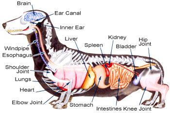
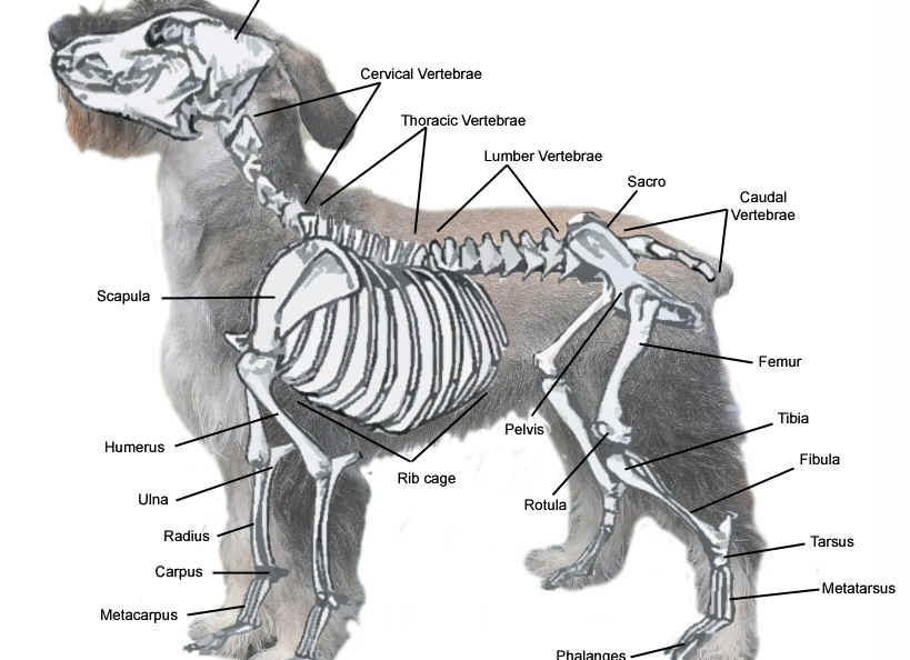
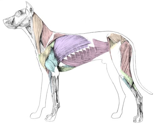
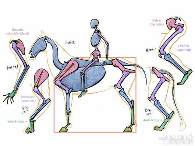
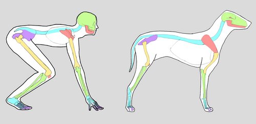
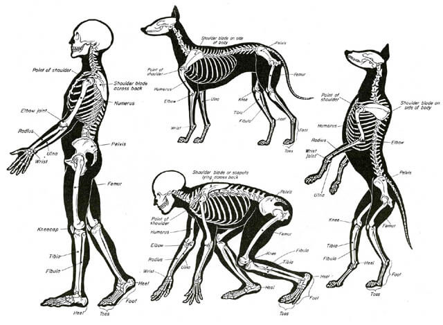
There are an enormous amount of traits that humans and animals share - this is because of the evolutionary process of inheriting characteristics and traits from successive generations that all lead back to a common ancestor. Humans and animals share the same basic muscles and bones, but they appear at different sizes, proportion and ratios based on the animal.
Bipeds are animals that traverse their environment on two legs, like us humans. Quadrupeds are animals that use four limbs to travel around like dogs, horses, cows, cats, and many other four limbed mammals. In terms of locomotion, evolution has developed two very common forms of movement using the same muscles and bones. As shown below, humans and dogs share the same groups of bones and muscles even though they have completely different forms of locomotion. In diagram A, a human man is shown next to a dog, the bones are highlighted on each animal and they are shown to be the same bone but in different proportions and ratios. What many people would think to be a dog's upper leg is actually its lower leg, and what many people think is it slower leg is actually the equivalent of a human palm.
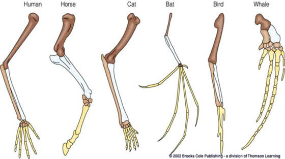
Using the same two animals as a comparison, human hands and dog paws when seen side by side share the exact same bones in different places. As seen in diagram B the thumb of the human is a vestigial part of the dog's foot, meaning a mutation from a previous ancestor that still appears in subsequent generations but is no longer used for the same purpose. In the comparison shown below in diagram C, the same bones shared between humans, large cats, and horses are pointed out, it is clear that many mammals have very similar skeletal structures regardless of their form of locomotion. Like the common misconception about dogs, the upper leg of most quadrupeds is hidden behind layers of muscle and fat, this is why colour coded Skelton diagrams are the most digestible forms of delivering information about the similarities and differences between human and animal anatomy.
Another very interesting area of anatomy that shares similarities and differences across multiple different species types is the bones of the hand. The human hand can be seen in many other animals such as bats, birds whales, horses, cats and other mammals. The diagram below shows how the same bones are reconfigured in other species to suit different purpose, including completely different types of locomotion including deep sea diving and swimming and even flight. It is interesting to see how the bones that we would see as the fingers can be fused together to create bird wings, or they can splay out to create bat wings. In the example of the horse, the "foot" of the horse that the hoof appears on is actually just one "finger" bone.
DOG vs CAT ANATOMY This material proudly presented by WWW.DOGICA.COM
Feline Paw Anatomy Feline front paws have a total of seven pads. There are five digital pads, one large plantar pad (heel pad) and one small wrist pad. Feline back paws have a total of five pads. There are four digital pads and one large plantar pad (heel pad). Typically felines have five nails on the front paws, one each to the digital pad. Due to a genetic disorder, some felines may have up to seven nails. Felines have four nails on the back paws, one to each digital pad.
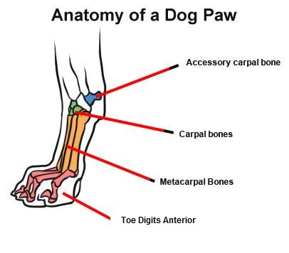
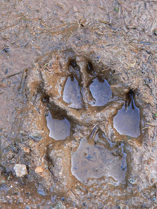
Canine Paw Anatomy Canine front paws have a total of six pads. There are four digital pads, one large metacarpal pad (heel pad) and one carpal pad. Canine back paws have five pads.
There are four digital pads and one large metacarpal pad (heel pad). Canines have five nails on the front paws, one each to the digital pad. Canines have four nails on the back paws, one each to the digital pad. Sometimes canines have a fifth nail higher up on the paw called the dew claw and in many instances this is removed when young.
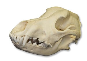
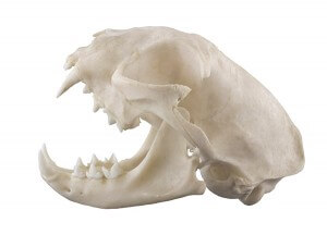
Dog vs Cat Skull Teeth Anatomy
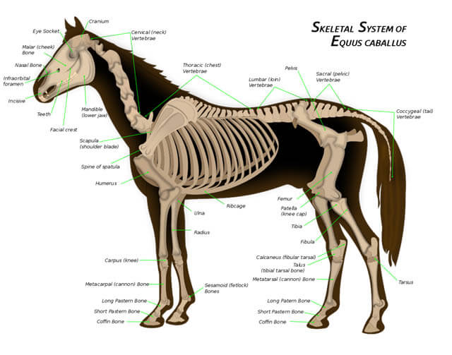
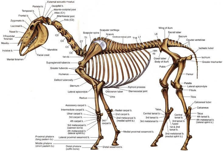
DOG AND HORSE ANATOMY: DIFFERENCES & SIMILARITIES This article is proudly presented by Erica Liszewski
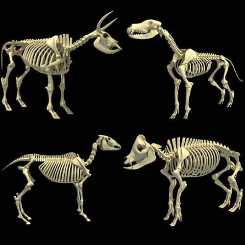
The basic structure of mammals, birds, reptiles and amphibians is very similar. We will provide both a carnivore (dog) and an herbivore (horse) in order to highlight some of the differences between them.
The dog and horse are probably the most well-studied of animals, so there is a lot of information available on both. I am also the most familiar with dog and horse anatomy, having drawn, studied, owned, and worked with both for many years. I'll start with the whole skeleton, and then look at each part in detail.
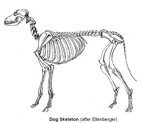
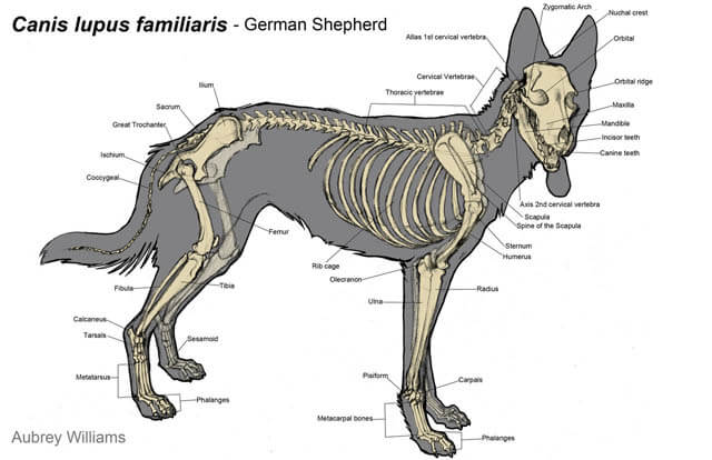
THE DOG SKELETON The dog is a carnivore, and while its evolution is not as specialized than the horse, the dog is constructed around teeth. The teeth of a dog are its primary source of food and defense. The dog has long thin limbs for running, although they don't need to travel as far or as fast as the horse.
Dogs have four "weight-bearing" digits per foot, with a shorter dew claw on the front and sometimes back feet. It is interesting to note that all domestic dogs have the same anatomy, the differences between breeds are in the size and shape of the bones.
THE HORSE SKELETON The horse is an herbivore, and its evolution has been driven by flight. A horse's life depends primarily on its ability to outrun danger. The horse has a shorter humerus and femur (upper leg), and a longer cannon bone (lower leg). This allows the horse to run faster, as it puts the muscle mass, and thus more of the animal's weight, closer to the body. The horse has only a single digit on each toe, although there are remnants of additional digits. Although the horse is more specialized than the dog, its structure is somewhat simpler.
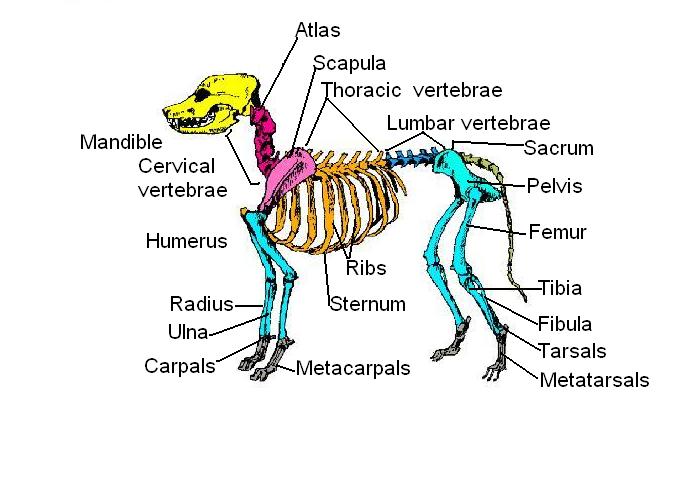
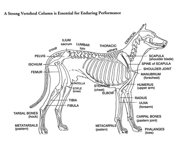
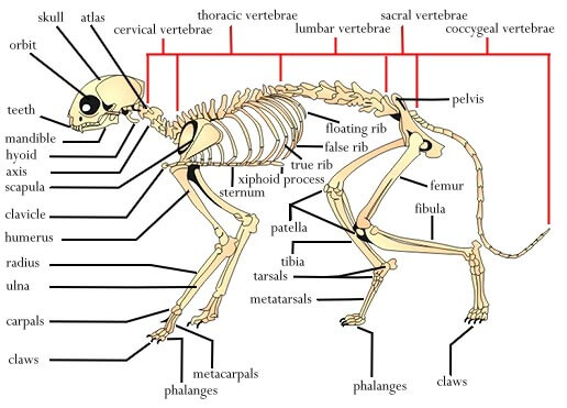
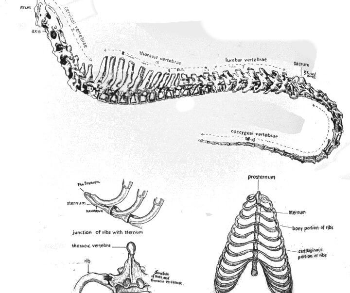
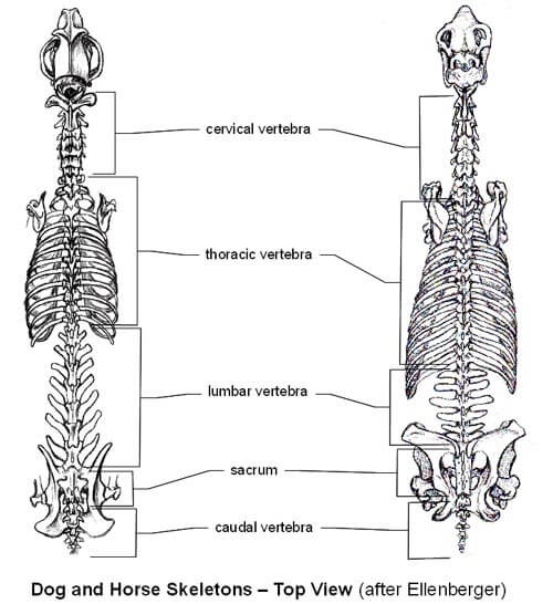
THE BODY Quadrupeds walk on all four limbs, and their bodies are built differently than bipedal animals. The spine of a quadruped contains the same types of vertebra as a human: cervical vertebra make up the neck, thoracic vertebra connect to ribs to form the chest, lumbar vertebra form the lower back, the sacrum attaches to the pelvis, and caudal vertebra form the tail. The exact number of each type of vertebrae varies somewhat between animals, but differences in shape are usually due more to the shape/size of vertebrae rather than a vast difference in number. Cervical vertebra are very flexible, and the first two are differently shaped to allow the head to pivot on the spine. Thoracic vertebra are much less flexible as each thoracic vertebrae attaches to a pair of ribs. Lumbar vertebra are fairly flexible in carnivores, but fairly rigid in herbivores. Not all animals have caudal vertebra, but they are always very flexible.
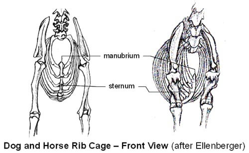
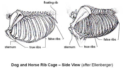
The rib cage of a quadruped is usually longer vertically - from sternum to spine, than across the body - from a given rib on the left to the corresponding rib on the right. The ribs spring out away from the spine at the top, and then curve back towards the mid-line towards the bottom. The forward sets of ribs connect directly to the sternum, and are called "true ribs". The next set of ribs are connected to the rib in front of them by cartilage, and are called "false ribs". Ribs that do not attach to any other are called "floating ribs". Carnivore ribs tend to be slender, while large herbivore ribs are very wide. The very front end of the sternum is called the manubrium, and marks the "point of the chest". The "point of the chest" is an important landmark, as it remains stationary regardless to the position of the head and neck.
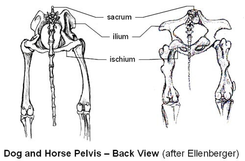
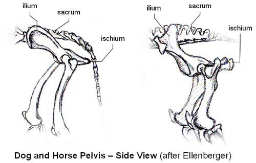
The pelvis is made up of mostly fused bones, and has little flexibility. Looking down the spine, the pelvis is ring shaped, with the top of the pelvis attaching to either side of the sacrum. From this ring, a "wing" called the ilium extends forward on each side. The shape of the ilium varies by species, but it tends to extend outwards more in herbivores than carnivores. The ends of this wing are a bony landmark called the "point of the hip" that is visible on most animals. Near the bottom, on either side of the "ring" are the sockets where the femur attaches to form the hip joint. The portion of the pelvis that extends backwards from the hip joint is called the ischium, which form the buttocks. The ischium is another bony landmark that is visible, or at least discernible, on most animals.
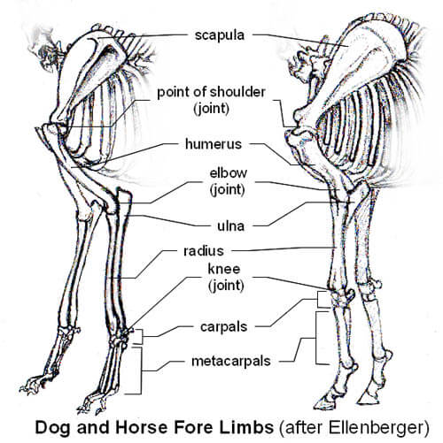
FORE LIMBS The fore limbs of the quadruped are weight bearing, and so differ from the fore limbs of a biped. The quadruped has an elongated scapula that lies alongside the rib cage, while the scapula of the biped lies behind the rib cage. The clavicle (collar bone) is usually small, or completely absent, and the fore limb is only attached to the body by muscle.
The scapula usually slopes forward and meets the humerus at the "point of the shoulder". The "point of the shoulder" is another useful landmark for animal anatomy. The humerus slopes backwards and meets the radius/ulna at the elbow. The radius and ulna are often fused in quadrupeds, which means they cannot rotate their "hands" like humans can. The elbow joint is usually close to the bottom of the rib cage, with the radius/ulna perpendicular to the ground. At the lower end of the radius/ulna is the "knee" joint - the human "wrist". This joint is usually higher - closer to the body in herbivores, and lower - closer to the ground, in carnivores.
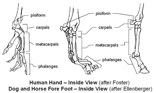
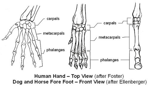
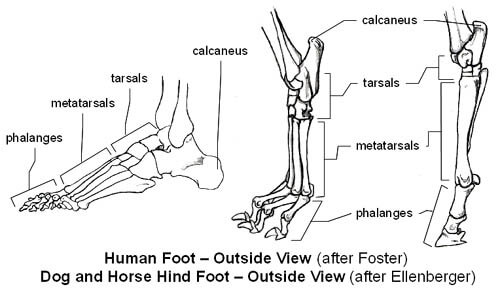
The knee joint itself contains two horizontal rows of small cube-shaped bones called carpal bones - the human wrist. The pisiform is the most obvious carpal bone, as it projects backwards from the joint. Below the knee are the metacarpals (hand bones), which are often fused in herbivores. Below the metacarpals are the phalanges, (finger bones), which may be split into toes (cow, deer), or remain a single toe (horse). Hoofed animals are literally walking on their fingertips. Carnivores have separate metacarpals, each ending a clawed toe. Carnivores walk on the last bone at the end of the "finger" with large pad supporting the weight from the "knuckle" joint.
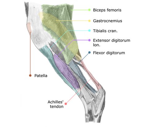
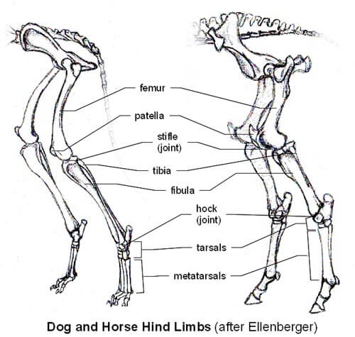
HIND LIMBS The hind limbs of a quadruped provide the power for movement. The femur (thigh bone) attaches to the pelvis with a ball and socket joint. The ball and socket joint allows for movement in multiple directions - inwards towards the mid-line, outwards, - away from the mid-line, as well as forwards and backwards. The lower end of femur is deeply notched in the front, were the patella kneecap fits and slides with the movement of the joint.
The tibia meets the femur at the stifle - the human "knee", which is usually just below the belly-level of a quadruped. The tibia is accompanied along it's length by the fibula, however the fibula is only partial in some animals - ox, horse. At the lower end of the tibia, and fibula if present, is the hock joint - the human "ankle". Similarly to the knee of the fore limb, the hock is usually higher on herbivores, and lower on carnivores.
The hock joint is made up of small tarsal bones, with the most prominent being the calcaneus (heel bone) which projects outwards and upwards from the back of the joint. The calcaneus bone is very obvious in most quadrupeds, as it will form the point at the back of the hock.
Below the hock are the metatarsals foot bones, which, like the metacarpals of the fore limb, are often fused in herbivores and separate in carnivores. The toes of the hind limb are very similar to the toes of fore limb, except that in carnivores the dew claw is usually absent. The hind feet of a quadruped will also generally be slightly narrower than the front feet, since most quadrupeds carry more of their weight on their fore limbs.
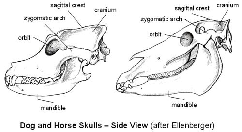
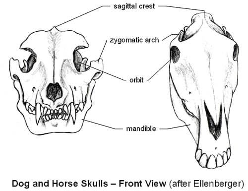
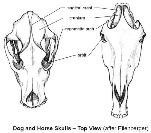
THE HEAD The skull of an animal serves to encase the brain, and houses the eyes, ears, nose, and mouth. The skull is usually only covered by small muscles, so the shape of the head is largely defined by the shape of the skull. The brain in housed in the rounded cranium, near the rear of the skull. Large carnivores will often have a ridge along the top of the cranium, where the chewing muscles attach.
The orbits - eye sockets, are usually on the front of the head for a carnivore, providing better depth perception, and on the sides of the head for a herbivore, providing a wider area of visibility. The zygomatic arch forming the cheekbones, begins near the orbits and extends out away from the skull, then connects again near the back of the skull. When viewed from the top, the zygomatic arch is often the widest part of the skull.
The mandible - lower jaw is hinged near the back of the skull, meaning the entire lower jaw is involved when opening and closing the mouth. The upper end of the mandible passes through the space formed by the zygomatic arch. Teeth vary depending on function and age of the animal, but carnivore teeth are usually pointed, while herbivore teeth are comparatively flat. Some herbivores (ruminants) lack front teeth on their upper jaw.
WHEN THE ORGANS MEET... Hopefully you now understand a little more about how quadrupeds are put together. So, how do you go about using this new insight? Here are a few suggestions. Practice drawing animal skeletons. Pay special attention to joints, especially if they usually give you trouble. You don't have to use my illustrations, as there are plenty of better ones available, some specific resources are listed near the end of this article. Going to a museum and sketching skeletons can also be useful. When looking at animals - at the zoo, on screen, or even your pets try to figure out where various bones are. Where is the scapula, how does it move? How do the joints of the leg extend and flex? Where are the ribs?
This is particularly useful with a pet, where you can gently feel for some of the less obvious bones. When drawing animals, pay attention to where the bones of that animal are. If you are having trouble getting a joint to look right, roughly sketch in the bones, and see if that helps you figure out how everything should fit together. When designing your own creatures, think about what their skeletal structure might look like. Is you creature a carnivore, herbivore, or omnivore? Does it depend on flight for survival, or does it fight? What type of terrain does it inhabit? How might these factors effect it is skeleton?
BY USING THIS SITE YOU ARE AGREE ON:
All materials on DOGICA® pages respectfully belong to its legal rights owners
All images on DOGICA® pages used only as illustrations. Find the author of any image with TINEYE
3rd Party cookies could be collected here by various installed widgets.
The information contained in or provided through DOGICA® site is intended for general consumer understanding and education only and is not intended to be and is not a substitute for professional advice.
Use of this site and any information contained on or provided through this site is provided on an "as is" basis without any representations, warranties or pay.
Consider disabling Ad Block in your browser to use Language Translator, Real-Time Visitors Map and Comments Box.